Our research
We develop quantitative MRI methods that aim to provide more objective, reproducible, and clinically useful measurements of tissue and disease. Our research spans MR acquisition, reconstruction, signal modelling, and quantitative mapping, with a strong focus on translating methodological developments into practical imaging tools.
We work across clinical MRI systems at 1.5T, 3T, and 7T and collaborate closely with clinicians, engineers, and industry partners. Our goal is to develop imaging approaches that are robust across field strengths and applications and that can ultimately contribute to faster, more scalable healthcare solutions.
A central challenge in quantitative MRI is to separate information about tissue properties from confounding effects such as field inhomogeneity, fat, motion, and multiple tissue compartments. We develop acquisition and analysis strategies that exploit the information contained in the MR signal while making these measurements more robust and reproducible.
Rather than focusing on a single organ or disease, we develop methodological concepts that can be translated across applications including the brain, heart, liver, musculoskeletal system, and emerging areas such as women’s health.
One example: exploiting signal asymmetries
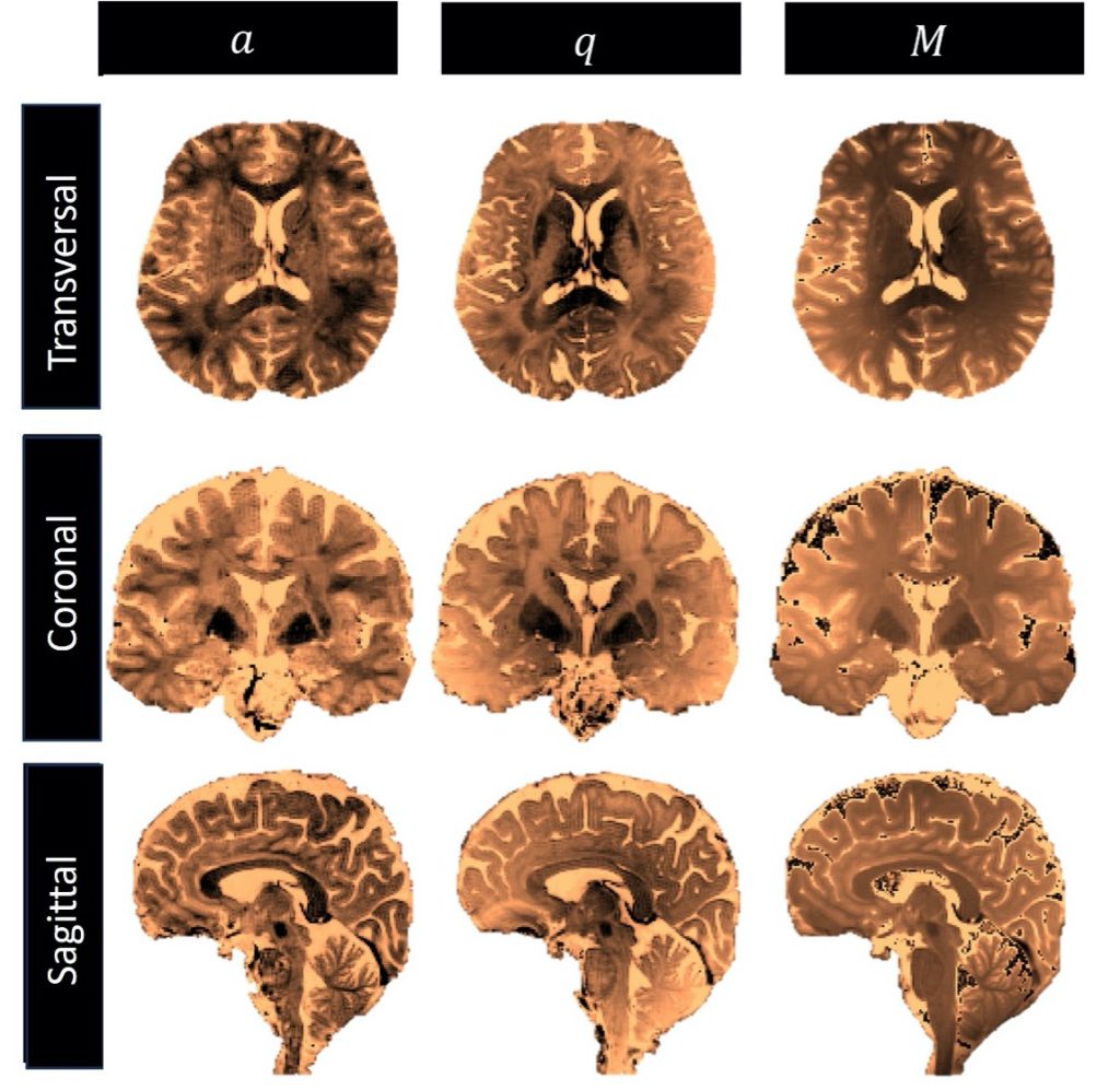
One example of our methodological research is phase-cycled balanced steady-state free precession (bSSFP). Instead of treating off-resonance-dependent signal variations simply as artifacts, we investigate how the structure of these signals can be used to encode additional quantitative information.
By combining MR physics, mathematical modelling, and efficient acquisition strategies, we use these signals to extract tissue parameters and develop new quantitative imaging approaches.
Our current research includes
- Multi-parametric quantitative MRI at 3T and 7T
- Robust imaging and quantitative mapping in the presence of off-resonance and field inhomogeneity
- Fat suppression and water–fat separation
- Motion-resolved cardiovascular MRI
- Quantitative imaging of the brain, heart, liver, and musculoskeletal system
- Open and vendor-independent MRI sequence development
- Integration of MRI with physiological and wearable sensor information
- New quantitative imaging approaches for women’s health
International and Industry Collaborations
Collaboration is central to how we work. We partner with clinical researchers, MR physicists, engineers, and industry to develop, validate, and translate new imaging technology.
We strongly support open science and the dissemination of methods beyond our own laboratory through open-source sequence implementations, shared software and data, and collaborations with research groups in Switzerland and internationally. We also work closely with Siemens Healthineers to translate selected methodological developments to clinical MRI systems.
Undergraduate and Graduate student projects
Students interested in MRI methodology, quantitative imaging, image reconstruction, computational modelling, or clinical translation are welcome to contact Prof. Jessica Bastiaansen. Projects are available for students with a wide range of backgrounds and levels of previous MRI experience.
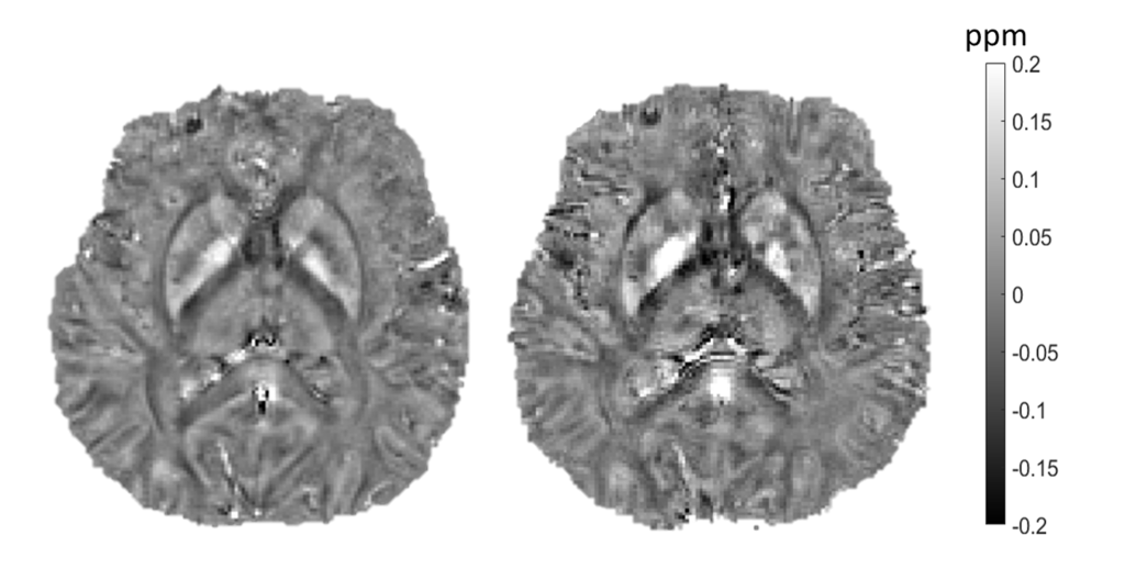
- Next-generation biomarker quantification in the human brain with innovative MRI techniques at ultra-high magnetic field.
- Quantification of brain microstructure dynamics using MRI
- MRI of the moving human eye at 7T
- Enabling direct imaging of neuronal activity with MRI
- Revolutionizing the assessment of cardiac function with cutting-edge Cine MRI techniques
- Motion-insensitive imaging of neuromuscular disease
- Novel methods for white and grey matter-specific biomarker quantification at 3T and 7T
- Advanced liver relaxometry to map fibrosis, fat and inflammation
- The influence of fat signal suppression on biomarker quantification with MRI
- Implementing and evaluating a vendor-independent 3D-radial sequence in an open-source framework
- Building a neural network for accurate quantitative MRI maps
- Mathematical modeling in MRI for multi-parameter estimation
- Solving MRI inversion problems for robust quantitative parameter extraction